Novogene AMEA
  • Novogene AMEA
  • Genomics
    • Human Whole Genome Sequencing
    • Plant and Animal Whole Genome Sequencing
    • Microbial Whole Genome Sequencing
    • Plant and Animal De novo Sequencing
    • Microbial De novo Sequencing
    • Shotgun Metagenomics Sequencing
    • Amplicon Sequencing
    • Whole Exome Sequencing
    Transcriptomics
    • mRNA Sequencing
    • Total RNA Sequencing
    • Full-Length Transcriptome Sequencing
    • Whole Transcriptome Sequencing
    • Small RNA Sequencing
    • Circular RNA Sequencing
    • Metatranscriptome Sequencing
    • Prokaryotic RNA Sequencing
    Single Cell & Spatial Omics
    • Single Cell Gene Expression
    • Single Cell Immune Profiling Sequencing
    • Single Cell Long Read Transcriptome
    • Visium HD Spatial Gene Expression
    • Stereo-Seq Spatial Gene Expression
    • Xenium In Situ Spatial Transcriptome
    Epigenomics
    • Whole Genome Bisulfite Sequencing (WGBS)
    • Directed DNA Methylation Sequencing (DM-Seq) NEW
    • Reduced Representation Bisulfite Sequencing (RRBS)
    • Chromatin Immunoprecipitation Sequencing (ChIP-seq)
    • RNA Immunoprecipitation Sequencing (RIP-seq)
    • Assay for Transposase-Accessible Chromatin with Sequencing (ATAC-seq)

    Premade Library

    • Sequencing Only on Illumina Sequencer
    • Sequencing Only on PacBio Sequencer
    Proteomics and Metabolomics
    • Olink Proteomics
    • Quantitative Proteomics
    • Untargeted Metabolomics
  • PromotionsPromotions
    • Platforms
    • Automated Delivery Platform (Falcon)
    • Bioinformatics Analysis Tool (NovoMagic)
    • Customer Service System (CSS)
    • Brochures
    • Case Studies
    • Webinar
    • Blog
    • Sample Guidelines
    • Cancer Research
    • Immuno-oncology
    • Agrigenomics
    • Environment
    • Food Science
    • Human Microbiome
    • Plant and Animal Microbiome
    • Drug Discovery and Development
    • Rare and Complex Diseases
    • About Us
    • Our Locations
    • News
    • Careers
  • Contact UsContact Us

ServicesServices menu

SupportSupport menu

CompanyCompany menu

Services
Whole Genome SequencingDe novo SequencingAmplicon SequencingShotgun Metagenomic SequencingDirected DNA Methylation Sequencing (DM-Seq)mRNA SequencingSingle Cell Gene ExpressionVisium HD Spatial Gene ExpressionXenium In Situ Spatial TranscriptomeOlink ProteomicsUntargeted Metabolomics
Support
NovoMagic Bioinformatics Analysis ToolCustomer Service SystemFalcon Intelligent Delivery Platform
Company
About UsOur LocationsOur PlatformsNewsCareersContact Us
LinkedInLinkedIn hoverYouTubeYouTube hoverXX hover
Copyright © 2026 Novogene Inc. All rights reserved.For Research Use Only. Not for Clinical Diagnostic Use.
Novogene AMEA
  • Novogene AMEA
  • Genomics
    • Human Whole Genome Sequencing
    • Plant and Animal Whole Genome Sequencing
    • Microbial Whole Genome Sequencing
    • Plant and Animal De novo Sequencing
    • Microbial De novo Sequencing
    • Shotgun Metagenomics Sequencing
    • Amplicon Sequencing
    • Whole Exome Sequencing
    Transcriptomics
    • mRNA Sequencing
    • Total RNA Sequencing
    • Full-Length Transcriptome Sequencing
    • Whole Transcriptome Sequencing
    • Small RNA Sequencing
    • Circular RNA Sequencing
    • Metatranscriptome Sequencing
    • Prokaryotic RNA Sequencing
    Single Cell & Spatial Omics
    • Single Cell Gene Expression
    • Single Cell Immune Profiling Sequencing
    • Single Cell Long Read Transcriptome
    • Visium HD Spatial Gene Expression
    • Stereo-Seq Spatial Gene Expression
    • Xenium In Situ Spatial Transcriptome
    Epigenomics
    • Whole Genome Bisulfite Sequencing (WGBS)
    • Directed DNA Methylation Sequencing (DM-Seq) NEW
    • Reduced Representation Bisulfite Sequencing (RRBS)
    • Chromatin Immunoprecipitation Sequencing (ChIP-seq)
    • RNA Immunoprecipitation Sequencing (RIP-seq)
    • Assay for Transposase-Accessible Chromatin with Sequencing (ATAC-seq)

    Premade Library

    • Sequencing Only on Illumina Sequencer
    • Sequencing Only on PacBio Sequencer
    Proteomics and Metabolomics
    • Olink Proteomics
    • Quantitative Proteomics
    • Untargeted Metabolomics
  • PromotionsPromotions
    • Platforms
    • Automated Delivery Platform (Falcon)
    • Bioinformatics Analysis Tool (NovoMagic)
    • Customer Service System (CSS)
    • Brochures
    • Case Studies
    • Webinar
    • Blog
    • Sample Guidelines
    • Cancer Research
    • Immuno-oncology
    • Agrigenomics
    • Environment
    • Food Science
    • Human Microbiome
    • Plant and Animal Microbiome
    • Drug Discovery and Development
    • Rare and Complex Diseases
    • About Us
    • Our Locations
    • News
    • Careers
  • Contact UsContact Us

ServicesServices menu

SupportSupport menu

CompanyCompany menu

Services
Whole Genome SequencingDe novo SequencingAmplicon SequencingShotgun Metagenomic SequencingDirected DNA Methylation Sequencing (DM-Seq)mRNA SequencingSingle Cell Gene ExpressionVisium HD Spatial Gene ExpressionXenium In Situ Spatial TranscriptomeOlink ProteomicsUntargeted Metabolomics
Support
NovoMagic Bioinformatics Analysis ToolCustomer Service SystemFalcon Intelligent Delivery Platform
Company
About UsOur LocationsOur PlatformsNewsCareersContact Us
LinkedInLinkedIn hoverYouTubeYouTube hoverXX hover
Copyright © 2026 Novogene Inc. All rights reserved.For Research Use Only. Not for Clinical Diagnostic Use.
banner laptop bg
banner mobile bg

Xenium In Situ Spatial Transcriptome

Request a Quote
(Xenium In Situ Spatial Transcriptome)
Request a Quote
(Xenium In Situ Spatial Transcriptome)
OverviewOverview
BenefitsBenefits
ApplicationsApplications
SpecificationsSpecifications
ResourcesResources

Xenium In Situ platform allows for simultaneous expressions analysis of more than 5000 target genes at subcellular resolution. This technology combines single-molecule RNA hybridization with sophisticated optical technologies to enable precise detection and analysis of over 5,000 target genes within individual tissue sections. It is compatible with fresh frozen (OCT embedded) and FFPE tissues, providing an unmatched microscopic perspective for applications in tumor biology, neuroscience, immunology, and beyond.

Why Choose Novogene for Xenium Spatial Transcriptomics?

Single-Cell Resolution & Multimodal Segmentation

Membrane, nucleus, cytoplasm staining with AI-based segmentation for precise cell boundary identification.

Single-Cell Resolution & Multimodal Segmentation

Membrane, nucleus, cytoplasm staining with AI-based segmentation for precise cell boundary identification.

High-Fidelity Information

Direct in situ imaging without RNA permeabilization preserves spatial context and molecular integrity.

High-Fidelity Information

Direct in situ imaging without RNA permeabilization preserves spatial context and molecular integrity.

Personalized Gene Customization

Supports custom targets, splice variants, cross-species analysis, and xenograft detection

Personalized Gene Customization

Supports custom targets, splice variants, cross-species analysis, and xenograft detection

High-Fidelity Information

Optimized probes and amplification enabling detection of low-abundance genes in complex tissue environments.

High-Fidelity Information

Optimized probes and amplification enabling detection of low-abundance genes in complex tissue environments.

Why Choose Novogene for Xenium Spatial Transcriptomics?

Single-Cell Resolution & Multimodal Segmentation

Membrane, nucleus, cytoplasm staining with AI-based segmentation for precise cell boundary identification.

Single-Cell Resolution & Multimodal Segmentation

Membrane, nucleus, cytoplasm staining with AI-based segmentation for precise cell boundary identification.

High-Fidelity Information

Direct in situ imaging without RNA permeabilization preserves spatial context and molecular integrity.

High-Fidelity Information

Direct in situ imaging without RNA permeabilization preserves spatial context and molecular integrity.

Personalized Gene Customization

Supports custom targets, splice variants, cross-species analysis, and xenograft detection

Personalized Gene Customization

Supports custom targets, splice variants, cross-species analysis, and xenograft detection

High-Fidelity Information

Optimized probes and amplification enabling detection of low-abundance genes in complex tissue environments.

High-Fidelity Information

Optimized probes and amplification enabling detection of low-abundance genes in complex tissue environments.

Applications of Xenium In Situ Spatial Transcriptome Sequencing

Unlock comprehensive insights into human genetics for hereditary disease, cancer, and population studies.

Tumor Microenvironment Research

Characterize the molecular and cellular landscape of the tumor microenvironment.

Tumor Microenvironment Research

Characterize the molecular and cellular landscape of the tumor microenvironment.

Immunology

Map the spatial distribution and interactions of immune cells within tissues.

Immunology

Map the spatial distribution and interactions of immune cells within tissues.

Neuroscience

Investigate neural cell function and expression patterns at unprecedented resolution.

Neuroscience

Investigate neural cell function and expression patterns at unprecedented resolution.

Biomarker Discovery

Identify cell-specific biomarkers for disease diagnosis and therapy development.

Biomarker Discovery

Identify cell-specific biomarkers for disease diagnosis and therapy development.

Applications of Xenium In Situ Spatial Transcriptome Sequencing

Unlock comprehensive insights into human genetics for hereditary disease, cancer, and population studies.

Tumor Microenvironment Research

Characterize the molecular and cellular landscape of the tumor microenvironment.

Tumor Microenvironment Research

Characterize the molecular and cellular landscape of the tumor microenvironment.

Immunology

Map the spatial distribution and interactions of immune cells within tissues.

Immunology

Map the spatial distribution and interactions of immune cells within tissues.

Neuroscience

Investigate neural cell function and expression patterns at unprecedented resolution.

Neuroscience

Investigate neural cell function and expression patterns at unprecedented resolution.

Biomarker Discovery

Identify cell-specific biomarkers for disease diagnosis and therapy development.

Biomarker Discovery

Identify cell-specific biomarkers for disease diagnosis and therapy development.

Specifications

Sample Requirements

Sample amounts are listed for reference only. Download the Sample Requirements to learn more. For detailed information, please contact us with your customized requests.

Sample TypeAmountStorage ConditionsSample QCShipping
FFPE Tissue Block1 block, FFPE block must be
contained on plastic dehydrating box,
otherwise could not be installed on the
sectioning device
After embedding
store at 4°C,
protected from light
DV200% ≥ 30%4°C or Room
Temperature

Sample Requirements

Sample amounts are listed for reference only. Download the Sample Requirements to learn more. For detailed information, please contact us with your customized requests.

Sample TypeAmountStorage ConditionsSample QCShipping
FFPE Tissue Block1 block, FFPE block must be
contained on plastic dehydrating box,
otherwise could not be installed on the
sectioning device
After embedding
store at 4°C,
protected from light
DV200% ≥ 30%4°C or Room
Temperature

Sample Requirements

Sample amounts are listed for reference only. Download the Sample Requirements to learn more. For detailed information, please contact us with your customized requests.

Sample TypeAmountStorage ConditionsSample QCShipping
FFPE Tissue Block1 block, FFPE block must be
contained on plastic dehydrating box,
otherwise could not be installed on the
sectioning device
After embedding
store at 4°C,
protected from light
DV200% ≥ 30%4°C or Room
Temperature

Sample Requirements

Sample amounts are listed for reference only. Download the Sample Requirements to learn more. For detailed information, please contact us with your customized requests.

Sample TypeAmountStorage ConditionsSample QCShipping
FFPE Tissue Block1 block, FFPE block must be
contained on plastic dehydrating box,
otherwise could not be installed on the
sectioning device
After embedding
store at 4°C,
protected from light
DV200% ≥ 30%4°C or Room
Temperature

Specifications

Sample Requirements

Sample amounts are listed for reference only. Download the Sample Requirements to learn more. For detailed information, please contact us with your customized requests.

Sample TypeAmountStorage ConditionsSample QCShipping
FFPE Tissue Block1 block, FFPE block must be
contained on plastic dehydrating box,
otherwise could not be installed on the
sectioning device
After embedding
store at 4°C,
protected from light
DV200% ≥ 30%4°C or Room
Temperature

Sample Requirements

Sample amounts are listed for reference only. Download the Sample Requirements to learn more. For detailed information, please contact us with your customized requests.

Sample TypeAmountStorage ConditionsSample QCShipping
FFPE Tissue Block1 block, FFPE block must be
contained on plastic dehydrating box,
otherwise could not be installed on the
sectioning device
After embedding
store at 4°C,
protected from light
DV200% ≥ 30%4°C or Room
Temperature

Sample Requirements

Sample amounts are listed for reference only. Download the Sample Requirements to learn more. For detailed information, please contact us with your customized requests.

Sample TypeAmountStorage ConditionsSample QCShipping
FFPE Tissue Block1 block, FFPE block must be
contained on plastic dehydrating box,
otherwise could not be installed on the
sectioning device
After embedding
store at 4°C,
protected from light
DV200% ≥ 30%4°C or Room
Temperature

Sample Requirements

Sample amounts are listed for reference only. Download the Sample Requirements to learn more. For detailed information, please contact us with your customized requests.

Sample TypeAmountStorage ConditionsSample QCShipping
FFPE Tissue Block1 block, FFPE block must be
contained on plastic dehydrating box,
otherwise could not be installed on the
sectioning device
After embedding
store at 4°C,
protected from light
DV200% ≥ 30%4°C or Room
Temperature

Demo Results

Image
Image
1/1
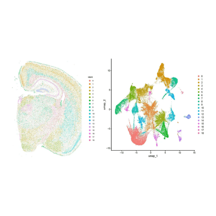
Cell Spatial Clustering Analysis: Spatial Distribution and UMAP

(Left) The spatial distribution of cells is shown, color-coded by graph-based clustering assignments. Each color represents a cluster of cells with similar gene expression profiles, providing insights into spatially distinct cell populations within the tissue. For large samples, only a subset of cells is visualized for performance optimization.

(Right) UMAP projection of cells, where each cluster is represented by a distinct color. This two-dimensional embedding highlights the relationships between clusters, with cells in closer proximity exhibiting more similar gene expression profiles. Subsets of cells are visualized for clarity in large datasets.

Image
Image
1/1
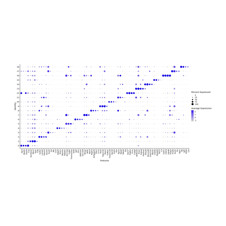
Bubble Plot for Marker Genes

The expression of marker genes in different clusters is calculated, and visualized as bubble plot. The results of the Top4 genes in each cluster are as above.

Image
Image
1/1
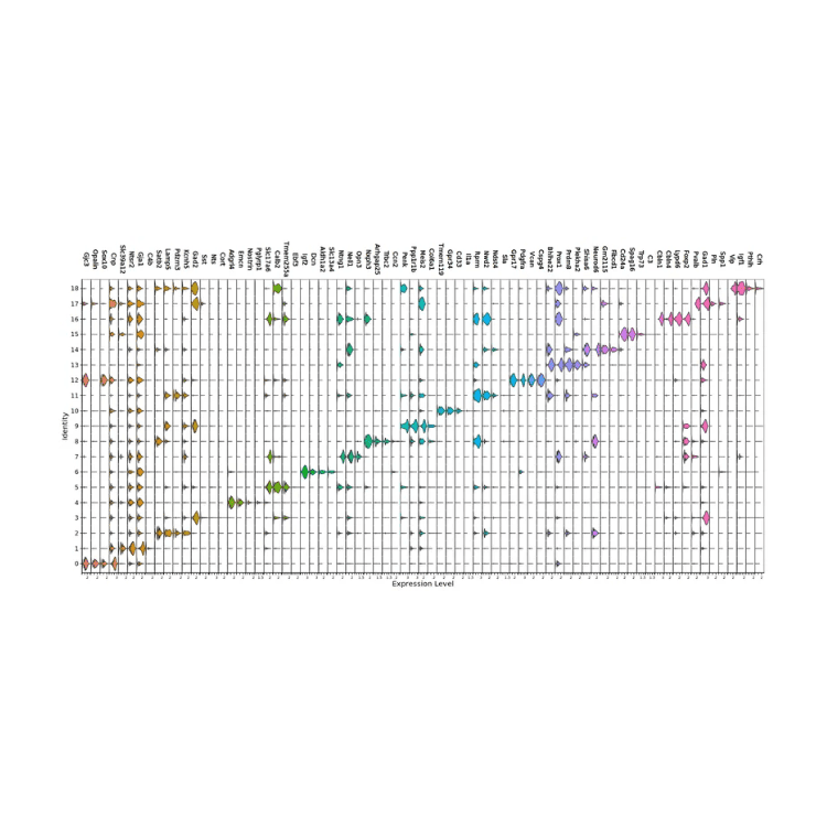
Marker Gene Violin Plot

The expression of marker genes in different clusters is calculated, and visualized as Violin Plot. The results of the Top4 genes in each cluster are as above.

Demo Results

Image
Image
1/1
Cell Spatial Clustering Analysis: Spatial Distribution and UMAP

(Left) The spatial distribution of cells is shown, color-coded by graph-based clustering assignments. Each color represents a cluster of cells with similar gene expression profiles, providing insights into spatially distinct cell populations within the tissue. For large samples, only a subset of cells is visualized for performance optimization.

(Right) UMAP projection of cells, where each cluster is represented by a distinct color. This two-dimensional embedding highlights the relationships between clusters, with cells in closer proximity exhibiting more similar gene expression profiles. Subsets of cells are visualized for clarity in large datasets.

Image
Image
1/1
Bubble Plot for Marker Genes

The expression of marker genes in different clusters is calculated, and visualized as bubble plot. The results of the Top4 genes in each cluster are as above.

Image
Image
1/1
Marker Gene Violin Plot

The expression of marker genes in different clusters is calculated, and visualized as Violin Plot. The results of the Top4 genes in each cluster are as above.

Image
Image
1/1
Cell Spatial Clustering Analysis: Spatial Distribution and UMAP

(Left) The spatial distribution of cells is shown, color-coded by graph-based clustering assignments. Each color represents a cluster of cells with similar gene expression profiles, providing insights into spatially distinct cell populations within the tissue. For large samples, only a subset of cells is visualized for performance optimization.

(Right) UMAP projection of cells, where each cluster is represented by a distinct color. This two-dimensional embedding highlights the relationships between clusters, with cells in closer proximity exhibiting more similar gene expression profiles. Subsets of cells are visualized for clarity in large datasets.

Image
Image
1/1
Bubble Plot for Marker Genes

The expression of marker genes in different clusters is calculated, and visualized as bubble plot. The results of the Top4 genes in each cluster are as above.

Image
Image
1/1
Marker Gene Violin Plot

The expression of marker genes in different clusters is calculated, and visualized as Violin Plot. The results of the Top4 genes in each cluster are as above.

More Services

Visium HD Spatial Gene Expression
(Visium HD Spatial Gene Expression)
Visium HD Spatial Gene Expression
(Visium HD Spatial Gene Expression)
Stereo-Seq Spatial Gene Expression
(Stereo-Seq Spatial Gene Expression)
Stereo-Seq Spatial Gene Expression
(Stereo-Seq Spatial Gene Expression)
Single Cell Immune Profiling Sequencing
(Single Cell Immune Profiling Sequencing)
Single Cell Immune Profiling Sequencing
(Single Cell Immune Profiling Sequencing)
Single Cell Gene Expression
(Single Cell Gene Expression)
Single Cell Gene Expression
(Single Cell Gene Expression)

More Services

Visium HD Spatial Gene Expression
(Visium HD Spatial Gene Expression)
Visium HD Spatial Gene Expression
(Visium HD Spatial Gene Expression)
Stereo-Seq Spatial Gene Expression
(Stereo-Seq Spatial Gene Expression)
Stereo-Seq Spatial Gene Expression
(Stereo-Seq Spatial Gene Expression)
Single Cell Immune Profiling Sequencing
(Single Cell Immune Profiling Sequencing)
Single Cell Immune Profiling Sequencing
(Single Cell Immune Profiling Sequencing)
Single Cell Gene Expression
(Single Cell Gene Expression)
Single Cell Gene Expression
(Single Cell Gene Expression)
Background
Background

Ready to Start Your Project?

Our platform offers tailored solutions for

your unique experimental needs, ensuring a seamless experience from project design to data delivery.


Privacy PolicyCookie PolicyCareers
banner laptop bg
banner mobile bg

Xenium In Situ Spatial Transcriptome

Request a Quote
(Xenium In Situ Spatial Transcriptome)
Request a Quote
(Xenium In Situ Spatial Transcriptome)
OverviewOverview
BenefitsBenefits
ApplicationsApplications
SpecificationsSpecifications
ResourcesResources

Xenium In Situ platform allows for simultaneous expressions analysis of more than 5000 target genes at subcellular resolution. This technology combines single-molecule RNA hybridization with sophisticated optical technologies to enable precise detection and analysis of over 5,000 target genes within individual tissue sections. It is compatible with fresh frozen (OCT embedded) and FFPE tissues, providing an unmatched microscopic perspective for applications in tumor biology, neuroscience, immunology, and beyond.

Why Choose Novogene for Xenium Spatial Transcriptomics?

Single-Cell Resolution & Multimodal Segmentation

Membrane, nucleus, cytoplasm staining with AI-based segmentation for precise cell boundary identification.

Single-Cell Resolution & Multimodal Segmentation

Membrane, nucleus, cytoplasm staining with AI-based segmentation for precise cell boundary identification.

High-Fidelity Information

Direct in situ imaging without RNA permeabilization preserves spatial context and molecular integrity.

High-Fidelity Information

Direct in situ imaging without RNA permeabilization preserves spatial context and molecular integrity.

Personalized Gene Customization

Supports custom targets, splice variants, cross-species analysis, and xenograft detection

Personalized Gene Customization

Supports custom targets, splice variants, cross-species analysis, and xenograft detection

High-Fidelity Information

Optimized probes and amplification enabling detection of low-abundance genes in complex tissue environments.

High-Fidelity Information

Optimized probes and amplification enabling detection of low-abundance genes in complex tissue environments.

Why Choose Novogene for Xenium Spatial Transcriptomics?

Single-Cell Resolution & Multimodal Segmentation

Membrane, nucleus, cytoplasm staining with AI-based segmentation for precise cell boundary identification.

Single-Cell Resolution & Multimodal Segmentation

Membrane, nucleus, cytoplasm staining with AI-based segmentation for precise cell boundary identification.

High-Fidelity Information

Direct in situ imaging without RNA permeabilization preserves spatial context and molecular integrity.

High-Fidelity Information

Direct in situ imaging without RNA permeabilization preserves spatial context and molecular integrity.

Personalized Gene Customization

Supports custom targets, splice variants, cross-species analysis, and xenograft detection

Personalized Gene Customization

Supports custom targets, splice variants, cross-species analysis, and xenograft detection

High-Fidelity Information

Optimized probes and amplification enabling detection of low-abundance genes in complex tissue environments.

High-Fidelity Information

Optimized probes and amplification enabling detection of low-abundance genes in complex tissue environments.

Applications of Xenium In Situ Spatial Transcriptome Sequencing

Unlock comprehensive insights into human genetics for hereditary disease, cancer, and population studies.

Tumor Microenvironment Research

Characterize the molecular and cellular landscape of the tumor microenvironment.

Tumor Microenvironment Research

Characterize the molecular and cellular landscape of the tumor microenvironment.

Immunology

Map the spatial distribution and interactions of immune cells within tissues.

Immunology

Map the spatial distribution and interactions of immune cells within tissues.

Neuroscience

Investigate neural cell function and expression patterns at unprecedented resolution.

Neuroscience

Investigate neural cell function and expression patterns at unprecedented resolution.

Biomarker Discovery

Identify cell-specific biomarkers for disease diagnosis and therapy development.

Biomarker Discovery

Identify cell-specific biomarkers for disease diagnosis and therapy development.

Applications of Xenium In Situ Spatial Transcriptome Sequencing

Unlock comprehensive insights into human genetics for hereditary disease, cancer, and population studies.

Tumor Microenvironment Research

Characterize the molecular and cellular landscape of the tumor microenvironment.

Tumor Microenvironment Research

Characterize the molecular and cellular landscape of the tumor microenvironment.

Immunology

Map the spatial distribution and interactions of immune cells within tissues.

Immunology

Map the spatial distribution and interactions of immune cells within tissues.

Neuroscience

Investigate neural cell function and expression patterns at unprecedented resolution.

Neuroscience

Investigate neural cell function and expression patterns at unprecedented resolution.

Biomarker Discovery

Identify cell-specific biomarkers for disease diagnosis and therapy development.

Biomarker Discovery

Identify cell-specific biomarkers for disease diagnosis and therapy development.

Specifications

Sample Requirements

Sample amounts are listed for reference only. Download the Sample Requirements to learn more. For detailed information, please contact us with your customized requests.

Sample TypeAmountStorage ConditionsSample QCShipping
FFPE Tissue Block1 block, FFPE block must be
contained on plastic dehydrating box,
otherwise could not be installed on the
sectioning device
After embedding
store at 4°C,
protected from light
DV200% ≥ 30%4°C or Room
Temperature

Sample Requirements

Sample amounts are listed for reference only. Download the Sample Requirements to learn more. For detailed information, please contact us with your customized requests.

Sample TypeAmountStorage ConditionsSample QCShipping
FFPE Tissue Block1 block, FFPE block must be
contained on plastic dehydrating box,
otherwise could not be installed on the
sectioning device
After embedding
store at 4°C,
protected from light
DV200% ≥ 30%4°C or Room
Temperature

Sample Requirements

Sample amounts are listed for reference only. Download the Sample Requirements to learn more. For detailed information, please contact us with your customized requests.

Sample TypeAmountStorage ConditionsSample QCShipping
FFPE Tissue Block1 block, FFPE block must be
contained on plastic dehydrating box,
otherwise could not be installed on the
sectioning device
After embedding
store at 4°C,
protected from light
DV200% ≥ 30%4°C or Room
Temperature

Sample Requirements

Sample amounts are listed for reference only. Download the Sample Requirements to learn more. For detailed information, please contact us with your customized requests.

Sample TypeAmountStorage ConditionsSample QCShipping
FFPE Tissue Block1 block, FFPE block must be
contained on plastic dehydrating box,
otherwise could not be installed on the
sectioning device
After embedding
store at 4°C,
protected from light
DV200% ≥ 30%4°C or Room
Temperature

Specifications

Sample Requirements

Sample amounts are listed for reference only. Download the Sample Requirements to learn more. For detailed information, please contact us with your customized requests.

Sample TypeAmountStorage ConditionsSample QCShipping
FFPE Tissue Block1 block, FFPE block must be
contained on plastic dehydrating box,
otherwise could not be installed on the
sectioning device
After embedding
store at 4°C,
protected from light
DV200% ≥ 30%4°C or Room
Temperature

Sample Requirements

Sample amounts are listed for reference only. Download the Sample Requirements to learn more. For detailed information, please contact us with your customized requests.

Sample TypeAmountStorage ConditionsSample QCShipping
FFPE Tissue Block1 block, FFPE block must be
contained on plastic dehydrating box,
otherwise could not be installed on the
sectioning device
After embedding
store at 4°C,
protected from light
DV200% ≥ 30%4°C or Room
Temperature

Sample Requirements

Sample amounts are listed for reference only. Download the Sample Requirements to learn more. For detailed information, please contact us with your customized requests.

Sample TypeAmountStorage ConditionsSample QCShipping
FFPE Tissue Block1 block, FFPE block must be
contained on plastic dehydrating box,
otherwise could not be installed on the
sectioning device
After embedding
store at 4°C,
protected from light
DV200% ≥ 30%4°C or Room
Temperature

Sample Requirements

Sample amounts are listed for reference only. Download the Sample Requirements to learn more. For detailed information, please contact us with your customized requests.

Sample TypeAmountStorage ConditionsSample QCShipping
FFPE Tissue Block1 block, FFPE block must be
contained on plastic dehydrating box,
otherwise could not be installed on the
sectioning device
After embedding
store at 4°C,
protected from light
DV200% ≥ 30%4°C or Room
Temperature

Demo Results

Image
Image
1/1
Cell Spatial Clustering Analysis: Spatial Distribution and UMAP

(Left) The spatial distribution of cells is shown, color-coded by graph-based clustering assignments. Each color represents a cluster of cells with similar gene expression profiles, providing insights into spatially distinct cell populations within the tissue. For large samples, only a subset of cells is visualized for performance optimization.

(Right) UMAP projection of cells, where each cluster is represented by a distinct color. This two-dimensional embedding highlights the relationships between clusters, with cells in closer proximity exhibiting more similar gene expression profiles. Subsets of cells are visualized for clarity in large datasets.

Image
Image
1/1
Bubble Plot for Marker Genes

The expression of marker genes in different clusters is calculated, and visualized as bubble plot. The results of the Top4 genes in each cluster are as above.

Image
Image
1/1
Marker Gene Violin Plot

The expression of marker genes in different clusters is calculated, and visualized as Violin Plot. The results of the Top4 genes in each cluster are as above.

Demo Results

Image
Image
1/1
Cell Spatial Clustering Analysis: Spatial Distribution and UMAP

(Left) The spatial distribution of cells is shown, color-coded by graph-based clustering assignments. Each color represents a cluster of cells with similar gene expression profiles, providing insights into spatially distinct cell populations within the tissue. For large samples, only a subset of cells is visualized for performance optimization.

(Right) UMAP projection of cells, where each cluster is represented by a distinct color. This two-dimensional embedding highlights the relationships between clusters, with cells in closer proximity exhibiting more similar gene expression profiles. Subsets of cells are visualized for clarity in large datasets.

Image
Image
1/1
Bubble Plot for Marker Genes

The expression of marker genes in different clusters is calculated, and visualized as bubble plot. The results of the Top4 genes in each cluster are as above.

Image
Image
1/1
Marker Gene Violin Plot

The expression of marker genes in different clusters is calculated, and visualized as Violin Plot. The results of the Top4 genes in each cluster are as above.

Image
Image
1/1
Cell Spatial Clustering Analysis: Spatial Distribution and UMAP

(Left) The spatial distribution of cells is shown, color-coded by graph-based clustering assignments. Each color represents a cluster of cells with similar gene expression profiles, providing insights into spatially distinct cell populations within the tissue. For large samples, only a subset of cells is visualized for performance optimization.

(Right) UMAP projection of cells, where each cluster is represented by a distinct color. This two-dimensional embedding highlights the relationships between clusters, with cells in closer proximity exhibiting more similar gene expression profiles. Subsets of cells are visualized for clarity in large datasets.

Image
Image
1/1
Bubble Plot for Marker Genes

The expression of marker genes in different clusters is calculated, and visualized as bubble plot. The results of the Top4 genes in each cluster are as above.

Image
Image
1/1
Marker Gene Violin Plot

The expression of marker genes in different clusters is calculated, and visualized as Violin Plot. The results of the Top4 genes in each cluster are as above.

More Services

Visium HD Spatial Gene Expression
(Visium HD Spatial Gene Expression)
Visium HD Spatial Gene Expression
(Visium HD Spatial Gene Expression)
Stereo-Seq Spatial Gene Expression
(Stereo-Seq Spatial Gene Expression)
Stereo-Seq Spatial Gene Expression
(Stereo-Seq Spatial Gene Expression)
Single Cell Immune Profiling Sequencing
(Single Cell Immune Profiling Sequencing)
Single Cell Immune Profiling Sequencing
(Single Cell Immune Profiling Sequencing)
Single Cell Gene Expression
(Single Cell Gene Expression)
Single Cell Gene Expression
(Single Cell Gene Expression)

More Services

Visium HD Spatial Gene Expression
(Visium HD Spatial Gene Expression)
Visium HD Spatial Gene Expression
(Visium HD Spatial Gene Expression)
Stereo-Seq Spatial Gene Expression
(Stereo-Seq Spatial Gene Expression)
Stereo-Seq Spatial Gene Expression
(Stereo-Seq Spatial Gene Expression)
Single Cell Immune Profiling Sequencing
(Single Cell Immune Profiling Sequencing)
Single Cell Immune Profiling Sequencing
(Single Cell Immune Profiling Sequencing)
Single Cell Gene Expression
(Single Cell Gene Expression)
Single Cell Gene Expression
(Single Cell Gene Expression)
Background
Background

Ready to Start Your Project?

Our platform offers tailored solutions for

your unique experimental needs, ensuring a seamless experience from project design to data delivery.


Privacy PolicyCookie PolicyCareers